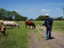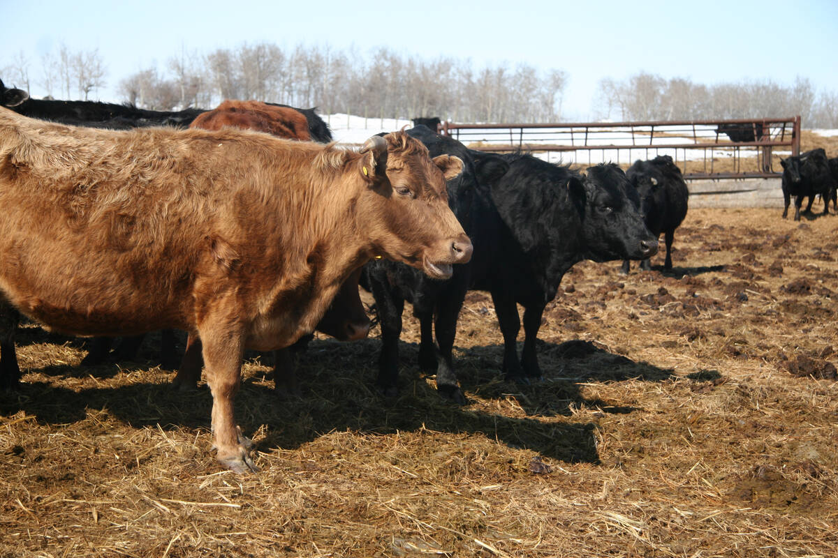
When pregnant cattle get infected with pathogens, there can be severe consequences for their fetal calves.
Bovine viral diarrhea virus (BVD) is an important example of this.
If the virus infects a cow, it spreads through the blood to the uterus, where it crosses the fetal membranes and infects the fetus.
Read Also
Canada’s simplified BSE testing program shows good uptake
Going by the number of submitted material samples so far, cattle producers’ response so far to an updated national surveillance program for BSE is encouraging for Canada’s CFIA.
This virus has a general preference for cells that are rapidly dividing. The area of the brain called the cerebellum is full of these rapidly dividing cells during fetal development and in the early neonatal period.
The BVD virus gains access to the fetal brain, likely from the blood vessels. When it arrives, it attacks dividing cells, including neurons and other cells within this small part of the brain. Because the cells die from the virus infection, there are no cells left to divide and grow this section of nervous tissue. The result is that the cerebellum is abnormally small.
Calves are then born with an underdeveloped cerebellum. This is the part of the brain responsible for regulating movement.
Calves with this abnormality are shaky, poorly co-ordinated and have difficulty walking. They stand with their legs widely spread to balance. Nursing can also be an issue for these calves.
The disease neither gets worse with time nor does it improve. Calves suspected of this condition are often euthanized, and the small cerebellum is confirmed during an autopsy examination of the brain. There are a few other viruses that may cause this, but they are far less common in Canadian cattle.
Another outcome of fetal viral infections in the brain is a condition called water on the brain, also known as hydrocephalus. Many different viruses can result in this disease.
Infection in the brain from these viruses kills the normal tissue that drains the fluid around the brain. As a result, the normal fluid movement in the brain is disrupted.
The excess fluid builds up, pushing the soft skull bones outward. Calves are born with a domed head and severe clinical signs such as seizures and mental dullness.
Severe hydrocephalus can also lead to difficult birthing because the deformed head is too large to pass through the cervix and vagina. These cows may need cesarean sections to get the calf out.
If the brain is dissected after death, it is reduced to a fluid-filled sac that collapses once the fluid drains because there is too little brain tissue left to support the structure.
Some viruses directly damage nerves responsible for movement, causing those portions of the nervous system to not develop properly.
The result is that fetal calves don’t move properly in the uterus. Their limbs are stiff and bent, which is called arthrogryposis. These calves may also have issues being born naturally.
The frustrating thing about these fetal infections is that they can be challenging to identify. The virus is often long gone by the time these calves are born. This means that our standard diagnostic tests to identify viruses won’t work.
In recent years, there has been work to uncover previously unknown viruses in cases of cattle brain disease. This is important work because the changes in a virus-infected brain are often not specific to any one virus. It takes additional molecular testing to uncover these causes.
With climate and other environmental changes, the distribution and timing of viral diseases are expected to change. Monitoring can help understand these changing dynamics.
Virus infections in pregnant cows can severely damage the nervous system of their fetal calves. For this reason, it is important to keep up farm infection control practices, including biosecurity measures and the use of vaccines to prevent infections.
Dr. Jamie Rothenburger, DVM, MVetSc, PhD, DACVP, is a veterinarian who practices pathology and is an assistant professor at the University of Calgary’s Faculty of Veterinary Medicine. X: @JRothenburger