Do you get confused when your veterinarian starts talking about cow reproduction?
You are not alone. So that you can understand what’s going on with cows, the following is a lesson on female body parts and how they work together to change an egg to a living calf.
The logical starting point is the ovaries. These two almond-shaped structures produce and release eggs. They are also an important source of female hormones.
A cow is born with 60,000 to 80,000 eggs or ova and this is all it is ever going to have. Throughout its reproductive lifetime, follicles, which are fluid-filled structures, form around individual eggs. Follicles grow and then rupture, releasing their eggs.
Read Also
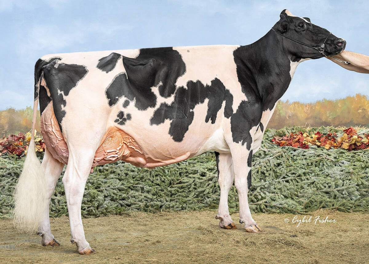
Saskatchewan dairy farm breeds international champion
A Saskatchewan bred cow made history at the 2025 World Dairy Expo in Madison, Wisconsin, when she was named grand champion in the five-year-old Holstein class.
Cells lining the growing follicle produce estrogen, a hormone that induces estrus behaviour. The cow emits an odour that attracts the bull and it becomes receptive to mating attempts. As the follicle reaches maximum size, there is a marked rise in the level of estrogen. This triggers rupture of the follicle, which we call ovulation.
Once the egg is released from its follicle, it flows into one of the two oviducts. The oviduct is like a funnel that surrounds an ovary and then narrows to a 25 centimetre long tube that connects to the uterus.
Minute, finger-like projections called cilia that line the oviduct keep the egg in constant motion so it doesn’t implant in the wall. The movement also promotes union of the egg and the sperm. Fertilization takes place in the oviduct.
The egg, fertilized or not, next moves down to the uterus. This is a bag-like structure designed to house the developing fetus. It has two horns, a relatively short body and is suspended in the abdomen by broad ligaments. Each oviduct empties into its own uterine horn.
After the egg is released, the follicular cells change to progesterone-producing cells. These are called luteal cells and the former follicle is called a corpus luteum.
Progesterone prepares the uterus for the fertilized egg. It causes blood vessels in the uterine wall to dilate and increases nutrient supply to the uterus. It also stops the rhythmic muscle contractions so the egg is implanted in the uterine wall.
The uterus also plays a role in assisting the sperm. Besides propelling the sperm toward the egg in the oviducts, secretions in the uterus help sperm capacitation, a maturation process that allows fertilization. The secretions also provide nourishment to the fertilized egg.
As the fetus grows, it covers itself with placental membranes, but, there is a connection between the dam and fetus. The umbilical cord leads to a placenta that has 70 to 120 exchange sites called caruncles. These allow nutrients from the mother to diffuse into the fetus and waste products to drain from the fetus to the dam.
The exit from the uterus is the vagina. Access to the vagina is controlled by a muscular ring called the cervix. This only opens at two specific times. It relaxes during estrus allowing sperm to gain entry, and it opens again during parturition to allow the calf to come out. Otherwise, it remains closed to prevent infection.
During parturition, muscles lining the uterus begin to contract, at first weakly, then as times goes on and the calf puts pressure on the cervix, with greater force. The calf is propelled through the cervix, enters the vagina and then passes out through the vulva. The vulva is the external opening through which pass urine and vaginal and uterine secretions, as well as the fetus and fetal membranes.