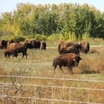
Three male calves died a few days after they were castrated and
vaccinated by the producer. Believing the deaths were linked to the
surgery, the attending veterinarian suspected that the calves succumbed
to tetanus as a result of infected castration sites.
This presumption turned out to be wrong.
The producer kept 32 cow-calf pairs on a brome-grass pasture, the same
land that his cows and calves had been allowed access to for several
years.
The three- to four-month-old calves were vaccinated with an intranasal
Read Also
Invigor Gold variety viewed as threat to condiment mustard
Invigor Gold, the canola-quality mustard developed by BASF, is on a collision course with Canada’s condiment mustard industry. It’s difficult to see how the two can co-exist.
IBR-PI3 vaccine and an injectable seven-way Clostridial,
bacterin-toxoid vaccine. The bull calves were castrated at the same
time as the vaccine administration.
They were castrated by cutting off the bottom of the scrotum and
removing the testicles with traction. They were pulled off rather than
cut off.
One calf died three days after processing. This was believed to be an
isolated incident, so no post mortem examination was done. Two days
later, a second calf died after showing symptoms of stiffness,
blindness and inco-ordination.
The veterinarian performed a post mortem
examination on the farm.
Because he saw no obvious lesions, he concluded that tetanus was the
cause of the death. Tetanus
antitoxin was given to the remaining calves as a
precaution.
A week later, a third calf was found down and kicking, but died before
the veterinarian arrived. A full post mortem was performed.
There was hemorrhage in the muscle of the heart and bleeding around the
bladder. Unfortunately, these lesions were not diagnostic for a
specific disease, so tissues were collected and submitted to the
laboratory for analysis. At this time, the differentials included acute
clostridial disease, tetanus, Haemophilus somnus and septicemia.
The investigation shifted when the pathologist was unable to grow
disease-causing infectious agents from the samples. A search for a
toxin was initiated.
The first analysis turned out to be the right one. Lead levels in the
calf’s blood were in the toxic range. With a level of 4.3 parts per
million, it was well over the limit of 0.4 ppm. The kidneys contained
101 ppm, well over the toxic level of 30 ppm.
Tracking down source
Once lead poisoning was diagnosed, the producer removed the cattle from
the pasture and began a search for a lead source. He was looking for
lead-
containing items such as old paint, linoleum, roofing felt, machinery
grease, caulking, arsenic defoliants and batteries.
An old lead-acid automobile battery that had been previously used for
an electric fence charger was soon discovered. After it was removed,
the calves and cows were allowed back on the pasture. No further
incidents of illness occurred.
The producer was worried that his cows, which were in early pregnancy,
could have been exposed to the lead, because it is known to cross the
placenta and enter the fetus. Luckily, the subsequent year’s crop did
not suffer unusual losses and the calves were born healthy.
The typical signs of lead toxicity are blindness, ataxia, depression,
episodes of running, bellowing, salivation, bloat and diarrhea. Nursing
calves are more likely to be affected because milk enhances lead
absorption. Young calves are inquisitive, which makes them more likely
to find a lead source and more likely to lick a novel object.
In this instance, it was difficult to make a diagnosis because the
veterinarian did not see live calves with clinical signs. A case like
this emphasizes the need for a complete postmortem when investigating a
death of unknown origin.
Jeff Grognet is a veterinarian and writer practising in Qualicum Beach,
B.C.