Fatal condition | Researchers are studying whether it is a different strain of the organism that infects cattle
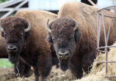
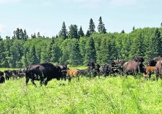
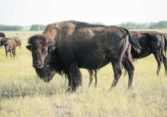
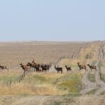
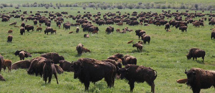
Mycoplasma bovis is a simple organism with devastating effects on bison.
Commonly known for causing pneumonia and arthritis in feedlot cattle, it is now found in domestic bison herds.
“The thing that is different and really surprised me was how many cases we discovered that was killing adult bison cows. We don’t see that in adult cattle,” said diagnostic pathologist Tom Clark, who is retired from the Western College of Veterinary Medicine.
“Something is amiss. Either the bison are extremely susceptible or we’ve got an organism that is quite active.”
Read Also
Feds propose overhaul of chronic wasting disease control program
Chronic Wasting disease control program getting updated by Canadian Food Inspection Agency with feedback encouraged from producers.
Mycoplasma bovis is considered a severe pathogen and is the most frequent cause of pneumonia, mastitis and arthritis in cattle.
Animals do not look sick until their lungs are almost destroyed. They do not appear to struggle to breathe but they may develop chronic laryngitis and become thin as the disease progresses. The calf is likely to die if a cow is infected because the mother cannot support it.
Veterinarian Pat Burrage of Bluffton, Alta., started a study after receiving calls from bison clients whose animals were getting sick in feedlots. Mycoplasma was diagnosed as the leading cause of death among these animals younger than 30 months.
A bigger problem emerged in adult animals older than 30 months.
“It seemed to us that it was a more significant condition in OTM (over 30 month) animals, meaning the cow-calf herds,” he said at a recent University of Calgary veterinary medicine conference.
Necropsies showed lesions were most consistent in the lungs, where they were nearly encased in whitish spongy abscesses, and chest cavities were full of smelly soup-like green fluid.
“The more I looked, the more I found when I started taking these animals apart,” he said.
He feared he had uncovered tuberculosis when he first started examining the dead animals. His study started in late 2010 and found 10 herds with at least one M. bovis related disease.
He worked with one producer who reported 22 abortions, 13 dead cows and eight to 10 chronically sick while another 30 were not affected.
“It is certainly a lot different in bison than it is in cattle,” he said. “It is something we didn’t see. Now we have uncovered the monster, we have to talk about it.”
More research is needed into where it came from and how it is spread, he added.
Clark said the organism is so small that large numbers can be expelled into the air through normal breathing and coughing.
“If they are in close contact, they are susceptible.”
The organism does not live outside the host for long so live animal contact is the likely cause. It could also travel in the wind.
“We know in equine diseases a virus can travel up to a kilometre in the wind,” said Burrage.
Researchers have not seen cross contamination on farms that raise more than one species. They want to know if this is the same bug infecting cattle or if they have encountered a new strain.
Mary Brown of the University of Florida, who studies mycoplasma in livestock, has also found it in alligators.
“We saw a situation where we had them die in 10 days,” she said.
Necropsies revealed arthritis, infection covering the lungs and enlarged hearts.
She suspects bison are so susceptible because they may not have been exposed to it in the past. It may take time for them to develop resistance.
“For right now, it is kind of scary seeing the parallels between the alligator and the bison.”
M. bovis is a simple organism surrounded by a cell membrane with lipid proteins that stick out from the surface. These can attach themselves to the host and feed off it.
It is sensitive to changes in the environment such as heat, dessication and most disinfectants. Antibiotics such as tetracyclines, fluorquinolones and macrolides will kill it. Penicillin does not work.
Some strains are more virulent than others. They are genetically distinct.
Even with DNA fingerprinting, researchers don’t know how relevant a specific genetic strain is to disease severity. It is possible to have 200 infected cows and 20 different isolates, but only two will cause serious disease, said Brown.
The classic way to diagnose it is with a culture, but it takes a long time to grow in the lab. This form of diagnosis is not practical if a feedlot has a suspected outbreak.
Further submissions may not show the disease if nose swabs were collected because it has lodged in the tonsils.
Scientists know it is a major worldwide pathogen, but it is primarily associated with intensely farmed beef and dairy cattle and calves.
“The clinical presentation will differ depending on the kind of husbandry and the type of operation,” she said. “If you have a group of animals with a high level of shedding then you are more likely to get rapid transmission.”
It may appear as a form of mastitis in dairy cows, which can periodically shed the bacteria and affect others.
Beef cattle may get pneumonia.
She looked at 359 dairy calves from birth and found that a disease spike showed up at about 50 days of age.
Animals did not show clinical disease once they were past weaning age and out on pasture.
She found they may appear if they can get past the early stages, but they may not thrive. They could have chronic respiratory disease, poor weight, head tilt and ear droop. The disease could spread to the central nervous system and they would be unlikely to recover.
Infected animals should be separated, and a veterinarian may recommend culling to prevent them from being carriers.
She also recommended using vaccinations for respiratory illness, improving hygiene and practicing good nutrition to give the animal a fighting chance.
In dairy animals, infection may come from direct contact with secretions in milk, nipples, buckets, tube feeders and bedding.