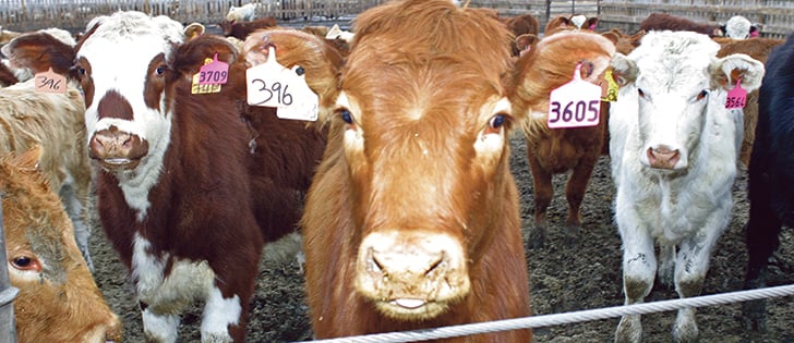

Cattle abortions can be frustrating for producers and veterinarians because a diagnosis may be confirmed only half the time.
“Getting negative findings from the lab does not equate with diagnostic failure,” said pathologist Cameron Knight of the University of Calgary’s faculty of veterinary medicine.
It simply means common causes were ruled out. The next step is to try to detect genetic abnormalities or environmental contaminants, he said at the university’s beef cattle conference in Calgary June 18-19.
A full-term calf could be a stillborn, while an abortion is a fetus that was expelled before term and would not be expected to live.
Read Also
Charges laid after cattle theft
Saskatchewan RCMP lay two charges against a man after six cattle went missing.
Early embryonic death is the hardest to diagnose. Cows return to estrus, an indicator of an early loss, but the cause may be unknown.
Tracebacks may find a bacterial infection, brucellosis, a virus such as bovine viral disease or a protozoa such as trichomoniasis or toxplasma.
Most producers become concerned when they notice three abortions within a short period of time. Knight offered a checklist of things to do and when to call the veterinarian.
- Save the specimens by bagging the fetus and/or placenta. Place them securely in a waterproof bag so they don’t leak, and put them in the refrigerator. Freezing tissues removes the ability to check for bacteria.
- Collect information because the vet will ask about the cow, feed supplies (such as moldy hay), gestation, climate, new animals in the herd and vaccination histories. Serial numbers are useful in case the vaccination failed.
- Examine the fetus. Few changes may be apparent, but it is wise to look for lesions or perhaps a congenital abnormality. An abnormality might be inside-out calves, two-headed calves or twins that did not form properly.
- Consider the gestational age of the fetus. This information is needed to make sure the calf was the right size for the expected stage of pregnancy. A bovine fetal growth chart can be downloaded from the internet to help estimate gestational age.
- Measure the top of the head to the pelvis and check to see how much hair had formed as further evidence of development.
- The state of decomposition should be noted. Some fetuses die slowly in the uterus, which could be the result of a maternal illness.
- Fetal intestines do not contain much material. The small amount of feces, called meconium, should not appear until after it is born. Animals that are distressed during birth will defecate and the body will be stained with its own feces.
- Examine the placenta. The afterbirth is often left in a mess in the calving area, but it can be the most useful tissue sample for diagnosis.
Veterinarians follow a standard procedure when checking an aborted fetus. Fresh samples are suitable for bacterial or fungal culture, while older samples are suitable for virology and toxins.
The lungs are checked during a necropsy to see if the animal drew breath to indicate whether it was born alive or was a stillbirth.
The veterinarian will also want a history of the dam and herd and may collect feed or water samples.