“There is nothing lazier than a cartilage cell; they just sit there,” quips a colleague in response to my comment on the beauty of these cells.
I’m looking through a microscope at a thin cross-section of a rat’s head, including brain, bone, teeth, and a stunning piece of cartilage.
I contemplate this comment.
We all know that the body is exquisitely complex with each cell contributing in its own unique way to the structure and function of the system.
But is it really fair to call cartilage lazy?
Read Also
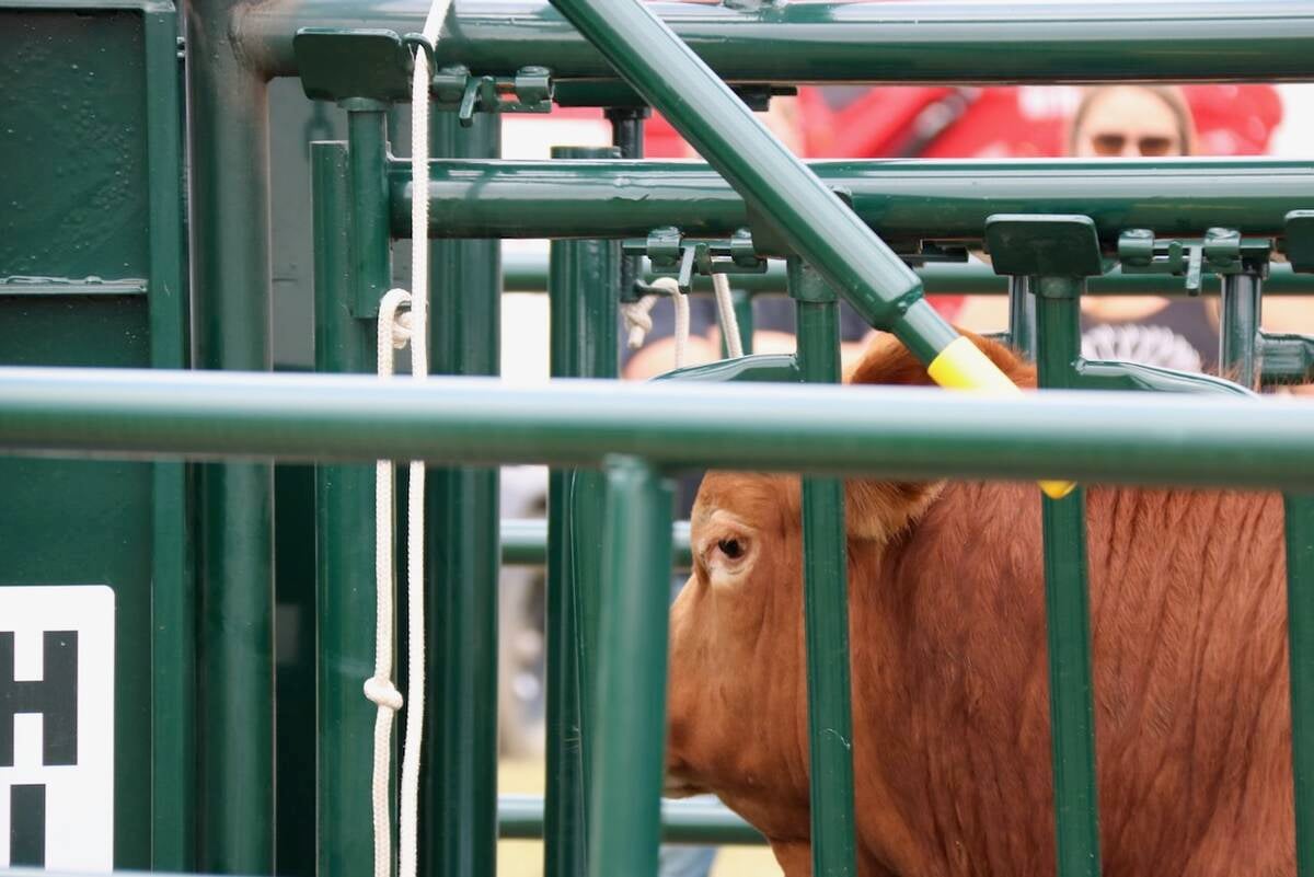
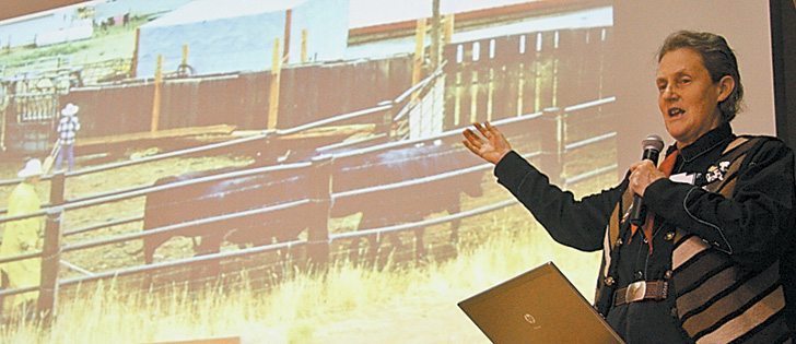
Good handling equipment a must on cattle operations
It’s important for the safety of producers and everyone else dealing with their stock that handling equipment is functional and safe.
In fetal development, a cartilage draft of the skeleton is made first. Bone is deposited onto this cartilaginous template.
Interestingly, sharks completely forgo this step of bone development and do just fine with a cartilage skeleton.
Cartilage is vital to the body’s structure from the beginning of an animal’s life. A body, like a house, needs a solid foundation.
Besides its role in the fetus, cartilage lines joints, allowing for freedom of movement by creating smooth surfaces on the ends of bones.
Cartilage is also found in unexpected places such as the airways, voice box, ear canal and intervertebral discs.
Cartilage doesn’t have a blood supply or nerve endings, yet these cells manage to secrete and maintain the collagen-rich material that provides strength and durability.
Surely it can’t be that lazy.
But cartilage can be injured.
Arthritis, which is inflammation of the joint, can occur in any animal. There are even signs of arthritis in dinosaur fossils.
Damage to the ligaments that support a joint creates unstable movement. This wears the cartilage unnecessarily, causing pain and limiting mobility.
The wear and tear that accompanies old age is another contributing factor in arthritis.
Old bulls and cows frequently have arthritis in multiple joints.
Cartilage doesn’t heal well because there is no direct blood supply and cartilage cells are slow to divide.
Then I consider the intestines.
The cells that line our digestive tract are the body’s extreme sport enthusiasts.
Despite their rapid division and death, these cells manage to do amazing tasks in their short lives, such as digesting food and absorbing nutrients and water from food while excreting wastes.
Specialized cells in the guts secrete mucous to smooth the way and protect from sharp food materials such as hay.
Horses are especially good mucous secretors.
In the face of viral infections, such as those that infect calves in the first few weeks of life, the intestinal lining cells are the first to be attacked and will often sacrifice themselves for the good of the body.
A virus’s ability to spread within the body is reduced when these cells die quickly and slough.
But what about the heart? Comprising specialized muscle cells, nerves, valves and connective tissue, these steadfast workhorses pump blood through a vast network of blood vessels, delivering oxygen and nutrients while removing wastes.
Consider the heart of an average sized cow.
At rest, it beats 60 times a minute and moves 35 litres of blood each time.
Unlike the intestines, heart cells cannot replicate themselves and must work tirelessly for the entire life of the body.
Broken hearts are difficult to mend because the cells cannot divide to replace those that are injured. Too many dropped beats and it is all over.
Bodies are much greater than the sum of their parts. Even though veterinarians focus on disease, it is often necessary to step back and remember how well the system generally works.
No matter how much or how little cartilage cells appear to do for the body as a whole, these cells provide an attractive microscopic view.