
Horses can develop cancer in their udders, which are their mammary glands.
It is a rare occurrence, with less than 20 cases reported in the scientific literature.
However, udder tumours in mares are serious. Most occur in middle-aged or older mares that are 12 to 25 years old.
There aren’t enough cases to identify other risk factors, such as breed or number of previous pregnancies, which may predispose mares to this type of cancer.
Udder tumours quickly grow into adjacent tissues and can spread to the lymph nodes and distant organs such as lungs, kidneys, ovaries, abdomen and chest cavity.
Read Also
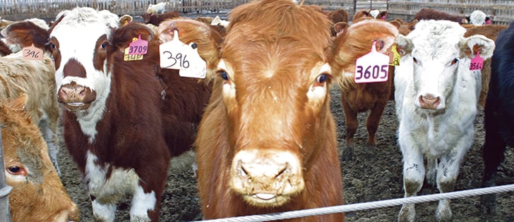
Charges laid after cattle theft
Saskatchewan RCMP lay two charges against a man after six cattle went missing.
Most mammary tumours originate in the udder, but sometimes tumours from elsewhere in the body can spread to the udder.
Clinical signs of mammary tumours in horses include edema that extends along the belly and sometimes down the back legs, swelling, pain, bleeding and ulceration of the skin on the udder.
Other systemic signs of cancer include weight loss, decreased appetite and stiff gait.
In most cases, both sides of the udder are affected, which poses a diagnostic challenge because it could be mistaken for mastitis, which is an infection and inflammation of the udder.
A key feature that may help distinguish cancer from mastitis is the presence of skin ulceration.
An incorrect diagnosis of mastitis would lead to unsuccessful treatment with antibiotics and pain medication, which would also delay potentially beneficial interventions such as surgery.
Veterinarians can confirm the cancer diagnosis with a needle biopsy and/or tissue biopsy in a living horse.
Sedation and freezing allows a veterinarian to insert a needle to collect cells, which are then examined microscopically.
Veterinarians could also remove a small amount of tissue from the affected udder and send it to a diagnostic laboratory.
With either technique, the predominant microscopic appearance is one of abnormal cancerous cells rather than pus, as would be expected with mastitis.
Horses are usually euthanized because of the severity of the disease, and the diagnosis is then confirmed with autopsy.
Carnivorous domestic animals, such as dogs and cats, are frequently diagnosed with mammary tumours with varying degrees of invasiveness.
However, horses are more likely than other livestock to receive intensive individual veterinary care and close monitoring while living to advanced ages, which means tumours in any part of the body, including the udder, have more opportunities to arise and be detected. Unlike dogs, mammary tumours in horses have a poor prognosis.
Treatment for udder tumours involves surgically removing the cancerous udder and lymph nodes in the area.
Chemotherapy is the mainstay for treatment of breast cancer in humans, dogs and cats, but the sheer size of horses makes treatment prohibitively expensive. Even with surgery, the cancer can return.
A common thread in these rare cases of udder tumours seems to be a delay in diagnosis.
The tumours are large and advanced before they are detected, which is easy to understand, considering it is an area where we don’t often look.
Horse owners and veterinarians should include an udder examination as part of their routine grooming and checkups.
Squat down and look at the udder from a safe distance. Gently feel both sides if the mare will tolerate it. Pay attention to swelling, asymmetries, pain, discoloration, ulcers and built up debris, and have them examined by a veterinarian if they persist.
If a mare is particularly sensitive, your veterinarian can do an udder exam when it is sedated for another procedure, such as after an annual dental examination and float.