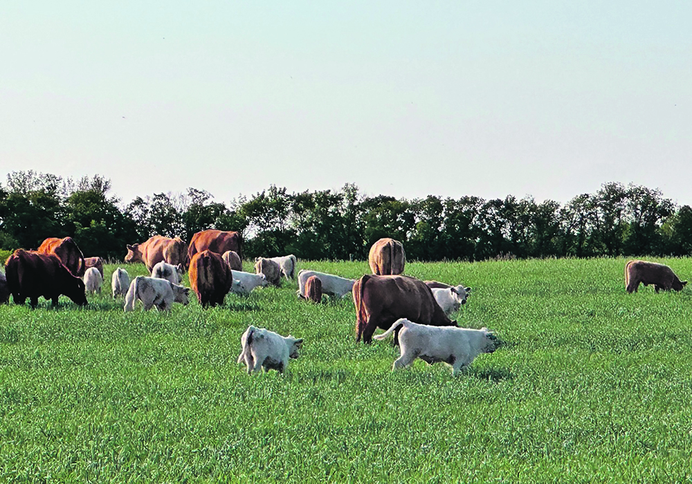
With spring and the births of calves, lambs, kid goats and foals nearly behind us, it is a good time to reflect on the amazing features of the placenta that make these little ones possible.
The placenta, along with the fetal membranes, forms what we typically call the afterbirth.
Not all mammals have placentas. Monotremes like the duck-billed platypus lay eggs. And there are examples of placentas and placenta-like structures in non-mammals, such as some sharks. But mammals with placentas are by far the most common group.
Read Also
Alberta cattle loan guarantee program gets 50 per cent increase
Alberta government comes to aid of beef industry with 50 per cent increase to loan guarantee program to help producers.
The placenta offers a distinct advantage over egg-producing animals. These include protection of the fetus in the internal environment of the dam, provision of high-quality nutrition and mobility. Egg-laying animals have to produce a relatively huge and energy expensive egg compared to animals with placentas, which may be less advantageous from an evolutionary perspective.
The fertilized egg develops into distinct layers quickly in embryonic development. It is the outer cell layer, called the trophoblast, that undergoes a series of co-ordinated, complex movements of cells and fusion of layers to eventually create the placenta. Although they differ in appearance between species and we can classify them in different ways, the basic function of the placenta remains the same between species.
Placentas are the important interface between the pregnant female and the fetus. It is intimately associated with the lining of the uterus and contains an intense network of blood vessels through which the fetus’s blood circulates.
These tiny blood vessels allow the exchange of molecules between the fetus and female. Gases (carbon dioxide and oxygen), nutrients (glucose sugars) and waste (excess nitrogen that will be excreted in the urine post-birth) all readily diffuse across. Those heavily pregnant cows and mares are breathing and eating for two and with goats and sheep often for three or more.
The blood vessels of the placenta converge to form the umbilical arteries and vein. These become thick, rope-like tubes in the umbilical cord that connects to the fetus at the navel.
In addition to their metabolic functions, placentas are a critical source of hormones. There is a complex interplay of hormones required for conception, pregnancy and birth of healthy offspring in animals.
It is the process of placental formation in most species that signals the pregnant female to stop cycling, in a process known as maternal recognition of pregnancy. The hormone progesterone is needed for successful pregnancy maintenance. And in horses and sheep, the placenta is the main source of this critical hormone for the last half of pregnancy. In cows, nanny goats and pigs, the ovary is the source throughout pregnancy.
Placentas protect the fetus by providing a barrier between it and the maternal system. The female’s immune system would attack the fetus as a foreign invader, if not for the placenta. Instead, it allows for a certain amount of tolerance for the fetus to develop. And the placenta is effective at keeping some toxins and infectious agents from attacking the fetus.
Some placentas support a single or at most, twin pregnancies, such as those in cattle. Others maintain huge litters such as those in pigs. Among common farm animal species, litter-producing pigs have the shortest pregnancies at approximately 114 days, while horses are the longest at 335 days.
The same hormones that allow birth to proceed are also necessary to expel the placenta. After the fetus is born, the placenta has to detach from the maternal tissues and is expelled from the uterus by similar uterine contractions, usually within a few hours following birth. Nearly all mammals consume some or all of the placenta. The reason for this behaviour is not completely understood but may be related to avoiding predation and taking advantage of its nutritional value.
Placentas play a remarkable role in animals.
In my next column, I’m going to discuss some things that can go wrong with this organ.
Dr. Jamie Rothenburger, DVM, MVetSc,PhD, DACVP, is a veterinarian who practices pathology and is an assistant professor at the University of Calgary’s Faculty of Veterinary Medicine. Twitter: @JRothenburger

