Q: I recently read your good article on skin cancer. It would be great to follow up with a colour photo of the three cancers. I used to have a chart that showed all three skin cancers. It is good to read about them, but pictures will save more lives.
A: A picture is worth a thousand words. I don’t usually add pictures to my column, but in the cases of skin problems, it is much easier to diagnose and recognize which condition a person has if you can see it.
Also, most skin cancers are first discovered by the individual and not the doctor.
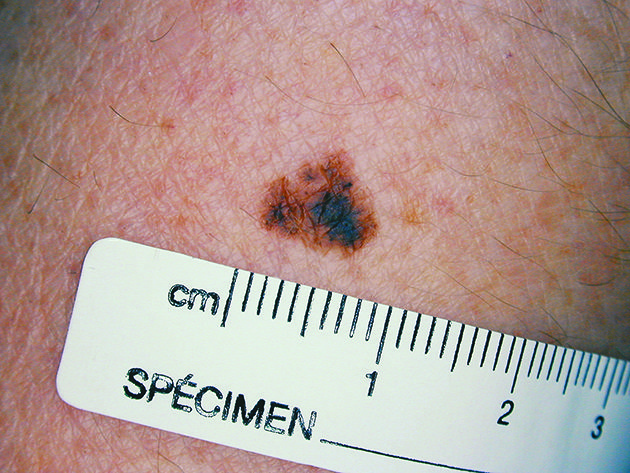
I will start with the most scary one, malignant melanoma, which can be fatal if not caught early. It develops from pigment cells in the skin known as melanocytes.
It usually does not develop from pre-existing moles, so there is no need to have regular moles removed as a precaution. It is dangerous because the cancer can spread to other parts of the body.
The edges of the mole are irregular and often reddish in colour. It looks as if it is spreading out into the surrounding tissue.
It can be raised and crusted, but not all crusty moles are malignant melanomas. Get it checked by your doctor.
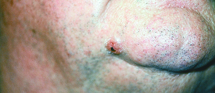
Basal cell carcinoma is often found on the nose or ears, a balding head or outsides of the hands because they are areas that get the most sun exposure.
It is often a small bump that does not start as a mole and is easily treated by surgical excision.
This is the most common type of skin cancer. Its precursor, actinic keratosis or solar keratosis, is caused by too much sun exposure.
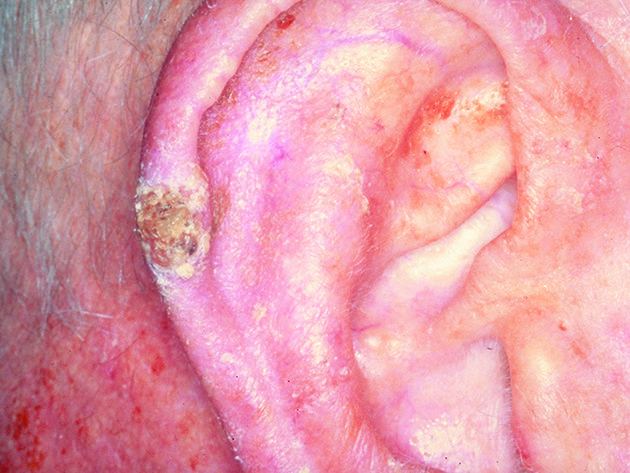
Squamous cell carcinoma, the second most common type of skin cancer, is treatable, but should not be ignored.
This variety can take several different forms. Early warning signs are a persistent scaly patch that can be reddish and irregular in shape. It can also be a raised area with a central depression that may sometimes bleed or an open sore that crusts and bleeds for weeks. They can look like warts, but warts do not bleed.