
Ever wonder about those little white spots in horses’ ears? They are called aural plaques, also known as ear papillomas. The white areas are where the skin is thickened due to infection with an equine papillomavirus.
Papillomaviruses, with and without associated skin changes, infect a wide range of species, including monkeys, dolphins, mice and cattle, as well as reptiles and birds.
In recent decades, there have been many new animal papillomaviruses discovered across a range of geographical locations and species. The diversity of this group of viruses closely follows the divisions between species.
Read Also
The Western Producer Livestock Report – October 2, 2025
Western Producer Livestock Report for October 2, 2025. See U.S. & Canadian hog prices, Canadian bison & lamb market data and sale insight.
It is likely that each species of animal has its own unique papillomavirus and that the host and virus evolved together over millions of years, making papillomaviruses among the oldest of viral families. It isn’t surprising that horses have a number of their own papillomaviruses.
Other species develop papillomavirus plaques similar to aural plaques in horses. I’ve diagnosed these on the skin of a wolf and they also occur in domestic dogs, in areas other than the ear.
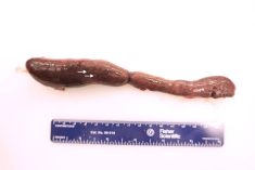
The lesions of aural plaques occur inside the outer pinna of the ear (the hair-covered pointy bits that are so expressive in horses, as opposed to the hidden inner ear). The affected skin has discrete white areas that are either flat or raised and between one and three centimetres in diameter.
While not distinct in light-coloured horses, these lesions can really stand out in the darker coats. The spots turn white because the virus infection causes the loss of normal skin pigment. In severe cases, the plaques can grow to be small, cauliflower-like masses that protrude above the skin surface like warts.
Under the microscope, affected ear skin is abnormally thickened by too many cells and too much of the outer flaky layer. The cells are packed with viruses, which are visible with high-powered electron microscopes.
Most affected horses have plaques in both ears but sometimes they occur only on one side. Aural plaques can occur in horses of any breed, colour, age or sex.
The ears of affected horses may have increased sensitivity, which can lead to handling issues for grooming, haltering, and bridling. Another negative impact is the presence of the plaques may decrease a horse’s economic value.
Papillomaviruses have a nifty, if sinister, ability to highjack the normal cell division process. The skin cells divide and grow much faster than normal when infected. They also keep the cell alive longer than normal to produce more viral copies. If we could give these viruses a trait, it would be safe to think of them as wanting skin cells to proliferate abundantly so that more virus is produced, which can be picked up by biting insects and passed on to other horses.
Any time the normal cell cycle is deranged, as it is with aural plaques, the possibility of cancer exists. However, with these ear masses, the transition to cancer is rare. There is one scientific report of a 28-year-old Thoroughbred gelding that had aural plaques and a common skin cancer called squamous cell carcinoma.
Currently, there is no commercial vaccine available to prevent infection and plaques don’t disappear on their own. Treatment with imiquimod cream was successful in a few published studies.
Successful treatment with this topical cream is intense and requires commitment. The ears need to be thoroughly cleaned before the cream is applied (usually requiring sedation), and this has to be repeated every two days. In one study, horses required eight to 30 treatments for complete resolution.
The virus that causes aural plaques is likely spread between horses by tiny black flies (Simulium spp.). Fly bites damage the skin, providing an entry point for the virus. They also likely act as a mechanical vector, carrying virus between horses, spreading the infection. For this reason, fly control can help limit spread.